PDR’s expert design engineers worked closely with the clinical lead on this project (Mr Cellan Thomas, Consultant Oral and Maxillofacial Surgeon, University Hospital of Wales) to plan the surgical procedure digitally, and design patient-specific devices. PDR’s carefully structured design process enabled efficient implementation of clinical decisions and contingencies in this time-sensitive project. One meeting was held to specify the surgical plan, with a second used to refine and approve the final device designs.
The Problem
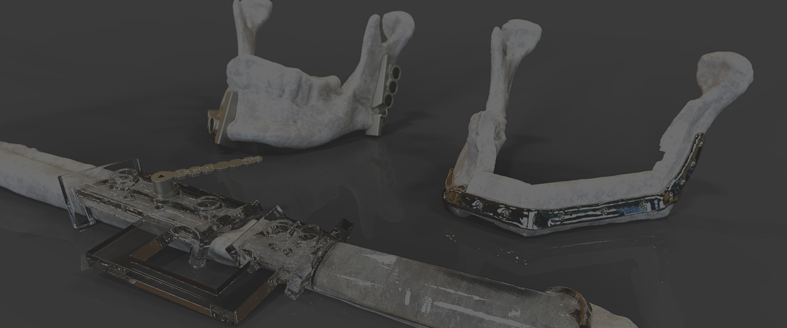
The patient had been diagnosed with a tumour in their lower jaw (mandible). The surgeon prescribed an excision with a safe margin; and replacement of the missing portion of jaw with carefully harvested and shaped segments of the patient’s fibula bone. This ‘fibula free-flap’ was selected because it had a good blood supply – which could be re-connected at the jaw-site.
Conventionally, this procedure would have relied heavily on an individual surgical team’s (considerable) skill on a given day. A digital design and 3d-printing approach was adopted to improve the predictability of the operation; and to reduce its duration - by eliminating processes which relied on iterative bone shaping and on-the-spot judgements. Ultimately, the aim was to guarantee superior aesthetic and functional results.
PDR segmented the patient’s CT scan-data and created a virtual model as the basis for digital surgical planning. The surgeon specified the cutting margins on the mandible, and the correct portions of fibula to reconstruct it. PDR designed patient-specific surgical guides to translate these cuts into the realities of theatre, and designed a custom implant to fit precisely against the bone segments and fix them together.
A third cutting guide was designed to ensure that exactly the correct lengths and angles of fibula bone were harvested. The cutting guides also featured drilling holes which precisely located pilot drills – and therefore the final screw-positions for fixing the implant. Drilling “keys” – developed and supported by PDR’s research – improved the accuracy of the drill guidance, and also encouraged good irrigation.
The implant, surgical guides, and PDR drilling keys were 3d-printed in medical-grade titanium under controlled conditions by our collaborators Renishaw PLC. The guides were post-processed to achieve a satin surface finish. Polishing achieved a smoother finish on the implant.
In theatre, all of the devices performed as pre-planned in the digital environment. Saw cutting vectors were translated exactly – and the correct portions of fibula harvested for the mandible. The guided pilot-holes combined well with the customised implant’s shape to quickly and accurately form the flap for transfer to the residual mandible.
The Result
Clinical post-operative assessment was positive. The position of the fibula segments was excellent – with analysis of the post-operative scans demonstrating excellent adherence to the surgical plan.


 Professor Dominic Eggbeer
Head of Surgical & Prosthetic Design
Professor Dominic Eggbeer
Head of Surgical & Prosthetic Design
 Surgical & Prosthetic Design
Enquiries & Orders
Surgical & Prosthetic Design
Enquiries & Orders
